Overview of Severs Disease
Severs disease, or calcaneal apophysitis, affects active children 8–15 years old, causing heel pain from growth plate inflammation. Conservative care—stretching, taping, heel lifts, and orthotics—helps reduce pain and maintain activity. Download PDF guides for home stretches and orthotic selection.
Common Symptoms and Clinical Presentation
Severs disease presents with a distinctive set of signs alerting clinicians and parents to growth‑plate inflammation at the calcaneus. Pain localizes to the posterior heel, described as a sharp ache that worsens with weight‑bearing activities like running or jumping. Parents note discomfort after play or school, and the child may refuse to bear weight. Pain often intensifies after rest and eases with gentle movement or stretching.
Pain may radiate into the calf, and swelling can be noticeable as a small bump at the heel base, mild soft
- Localized tenderness over the posterior heel and the calcaneal apophysis.
- Swelling or a small, firm mass at the heel base, sometimes visible as a bulge.
- Limited dorsiflexion of the ankle due to pain and stiffness.
- Visible guarding or limp when walking, especially on the affected side.
- Reduced tolerance for high‑impact sports, leading to decreased participation.
Physical exam shows a tender area above the heel bone, with pain when dorsiflexion or resisted plantar‑flexion. The child’s gait may have a subtle heel‑strike pattern, and calf muscles may feel tight. A normal knee and hip range is expected. These findings guide conservative care, including stretching, orthotics, and activity modification; During follow‑up, clinicians monitor resolution of swelling and pain, ensuring safe return to sport and adjusting orthotics as needed. Follow‑up essential.
Risk Factors and Epidemiology
Severs disease is most common in active children aged 8–15 years, coinciding with rapid growth spurts. The condition is strongly associated with high‑impact sports such as running, jumping, and soccer, where repetitive loading of the heel accelerates inflammation of the calcaneal growth plate. Boys and girls are equally affected, but boys may show earlier onset due to earlier growth spurts. Children who participate in competitive sports or engage in frequent high‑impact training have a higher incidence. Conversely, less active children can develop the disease, though at a lower rate. Other risk factors include a history of ankle sprains, tight calf muscles, and biomechanical abnormalities such as overpronation or high arches, which increase shear forces on the heel. Ethnic and genetic predispositions appear minimal; however, a family history of growth‑plate injuries may slightly elevate risk. Epidemiologic studies report prevalence rates ranging from 2% to 5% among school‑aged athletes, with peak incidence during the 12‑14 year age window. Early identification through routine screening in youth sports programs can reduce severity and improve outcomes.
In addition, nutritional deficiencies can impair bone mineralization, predisposing to growth‑plate stress. Children with flat feet have altered shock absorption, increasing heel loading. Overuse injuries from footwear or sudden increases in training volume also contribute. Screening for foot morphology and calf flexibility during growth spurts can identify at‑risk children early. Early intervention reduces long‑term complications soon. Kids heal fast.
Diagnostic Criteria and Imaging Findings
Severs disease is diagnosed clinically by a triad of heel pain, localized tenderness over the calcaneal apophysis, and pain that worsens with activity or weight‑bearing. The patient is typically an active child aged 8–15 years who has a recent growth spurt. A physical exam reveals pain on palpation of the posterior heel, swelling or erythema, and pain during resisted dorsiflexion or calf‑stretch maneuvers. The diagnosis is confirmed when these findings coexist with a normal gait pattern and the absence of systemic symptoms.
Imaging is reserved for atypical presentations or to rule out fractures. Plain radiographs show a widening or irregularity of the calcaneal apophysis, but are often normal in early disease. Ultrasound can demonstrate thickening of the calcaneal growth plate and increased vascularity, providing a non‑radiation alternative. MRI offers the most sensitive assessment, revealing bone marrow edema, soft‑tissue inflammation, and a hypointense rim around the growth plate. A typical MRI appearance includes a low‑signal calcaneal apophysis on T1 with high‑signal edema on T2/STIR sequences.
Diagnostic criteria integrate age, activity level, clinical signs, and imaging when necessary. Early recognition allows prompt conservative management, preventing progression to chronic heel pain or growth‑plate injury.
Differential diagnosis includes stress fractures, tarsal coalition, and plantar fasciitis. Radiographs may reveal a lucent zone or periosteal reaction, while ultrasound can detect periosteal fluid. MRI is preferred for early edema, showing a low‑signal zone on T1 and high‑signal on T2, confirming active inflammation. Early imaging prevents misdiagnosis and guides treatment.
These criteria help clinicians choose the right imaging and treatment plan. Soon.
Conservative Treatment Principles
Conservative management of Severs disease focuses on pain relief, inflammation control, and functional restoration while protecting the growth plate. Core principles include: 1) activity modification—temporarily reduce high‑impact activities such as running or jumping; 2) rest and ice—apply 15–20 minute ice packs every 2–3 hours during the first 48–72 hours to limit edema; 3) NSAIDs for short‑term use to reduce pain and inflammation, with careful monitoring for side effects; 4) structured calf‑stretching and strengthening—daily calf stretches for 30–45 seconds per side, combined with toe‑raiser and heel‑rise exercises to improve flexibility and reduce dorsiflexion load on the calcaneal apophysis; 5) orthotic support—custom or prefabricated heel lifts (1–2 cm) to off‑load the growth plate, and arch‑supporting insoles to maintain foot biomechanics; 6) taping—elastic or kinesiology tape applied in a dorsal‑to‑plantar direction to provide dynamic support; 7) progressive return to activity—gradual re‑introduction of sport drills once pain subsides, guided by pain thresholds and functional tests; and 8) patient education—informing children and parents about the growth‑plate nature of the injury, importance of adherence, and signs of worsening symptoms that warrant re‑evaluation. Consistent application of these principles typically results in pain resolution within 4–6 weeks and a safe return to athletic participation.
Physiotherapy sessions can incorporate manual therapy techniques such as gentle mobilization of the subtalar joint and soft‑tissue massage to improve circulation. Ultrasound‑guided injections of corticosteroids are rarely indicated but may be considered in refractory cases after thorough evaluation. Monitoring growth plate healing through periodic clinical reassessment and, if necessary, repeat imaging ensures that the intervention remains appropriate. The goal is to preserve the natural growth process while preventing chronic pain or deformity.
Calf Stretching Protocols for Severs Disease
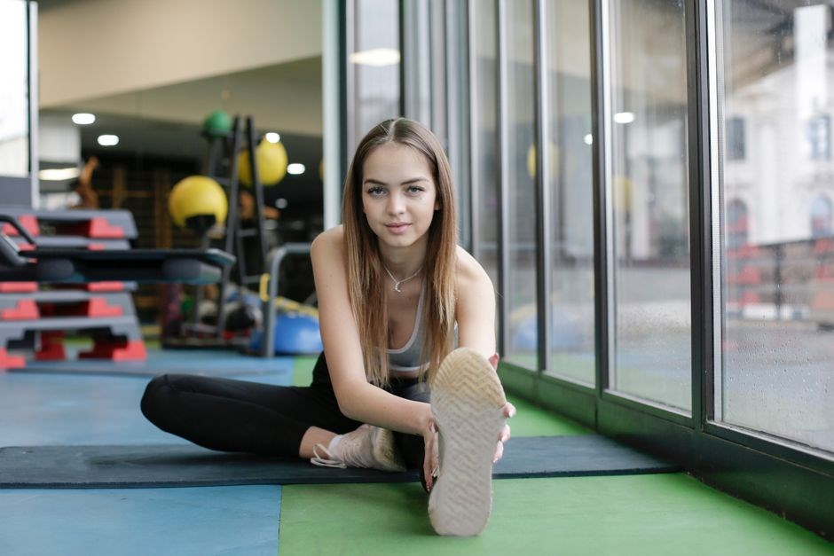
Effective calf stretching is the cornerstone of non‑operative care for Severs disease. The goal is to increase dorsiflexion range, reduce tension on the Achilles tendon, and a structured routine should be performed daily, twice a day, and can be divided into three phases: warm‑up, static stretch, and cool‑down.
Warm‑up (5–10 min)
Light walking or stationary cycling at a low intensity to elevate foot temperature and improve blood flow. This prepares the muscle for stretching and reduces the risk of strain.
Static stretch (30–45 s × 3–4 repetitions)
Wall calf stretch: Stand facing a wall, place both hands on the wall at shoulder height, and step the affected leg back keeping the heel flat. Lean forward until a gentle stretch is felt in the calf. Hold, then switch legs.
Seated toe‑raiser stretch: Sit on a chair, place the foot on a step or sturdy box, and slowly lower the heel below the step level. Hold the stretch for 30–45 seconds, then lift the heel. Repeat 3–4 times per leg.
Standing heel‑rise stretch: Stand on a flat surface and lift toes, lower heels, feeling stretch in Achilles.
Cool‑down (5 min)
Gentle ankle circles and light massage of the calf to promote circulation and aid recovery.
Progression: If pain is minimal, increase hold time to 60 seconds and add a second set of 3–4 repetitions. Avoid overstretching; pain should be mild and transient. Consistency is key—skipping sessions can delay healing and prolong discomfort. Document each session in a simple log to track adherence and pain changes.
These protocols are supported by systematic reviews indicating that regular calf stretching reduces pain scores and allows children to maintain participation in sports. For a printable PDF guide, refer to the downloadable resource linked below.
Download the PDF for guidance now here
Calf Strengthening Exercises
Strengthening the gastrocnemius and soleus helps off‑load the inflamed growth plate and improve dynamic stability during activity. A progressive, low‑impact routine can be performed at home or in clinic. The program is divided into three phases: initiation, progression, and maintenance.
Initiation (Weeks 1–2)
Toe‑raiser on a step: Stand on a step with heels hanging over the edge. Slowly raise the toes, hold for 2 seconds, then lower heels below step level. Perform 2 sets of 10 repetitions, twice daily.
Seated heel‑drop: Sit on a chair, place the foot on a step, and slowly lower the heel while keeping the toes on the step. Hold for 5 seconds, then lift back. 3 sets of 8 reps, twice daily.
Progression (Weeks 3–4)
Add resistance by holding a light dumbbell (1–2 lb) in the hand opposite the working leg. Increase repetitions to 12 per set and introduce unilateral calf raises on a low step. Perform 3 sets of 12, twice daily.
Maintenance (Weeks 5+)
Continue unilateral calf raises, increasing step height gradually. Perform 3 sets of 15 reps, twice daily. Incorporate plyometric drills (e.g., low‑impact hopping) permits.
Throughout the program, monitor pain; any sharp discomfort should halt the exercise. Consistency, gradual load increase, and proper form are key to preventing recurrence and ensuring a safe return to sport.
Download the PDF for a printable exercise sheet here.
Closed‑Chain Dorsiflexion Technique
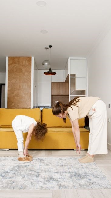
Closed‑chain dorsiflexion is a low‑impact exercise that mobilizes the ankle joint while keeping the foot planted, thereby reducing shear forces on the calcaneal growth plate. The movement is performed on a step or curb and focuses on gentle dorsiflexion with the knee aligned over the fourth toe.
Step 1 – Position the foot on a step so that the heel remains on the ground. Step 2 – Align the knee directly above the fourth toe, ensuring the knee does not collapse medially or laterally. Step 3 – Lean forward slowly, allowing the ankle to dorsiflex while the heel stays planted. Hold the position for 5 seconds, feeling a mild stretch in the calf and Achilles tendon.
Repeat the movement 10 times, then rest for 30 seconds. Perform two sets per session, twice daily. Gradually increase hold time to 7–10 seconds as tolerance improves. This technique promotes joint mobility, improves calf flexibility, and supports off‑loading of the inflamed growth plate.
For best results, combine the closed‑chain dorsiflexion with calf strengthening and taping protocols. A printable PDF guide is available for step‑by‑step instructions and visual cues.
Clinical guidance recommends performing the closed‑chain dorsiflexion with a metronome set to 2 seconds per repetition. Patients should monitor pain, stopping if discomfort exceeds a 3‑point threshold on a 0‑10 scale. Documentation of progress aids future adjustments. Use a light ankle brace for stability!!!.
Heel Lift Usage and Selection
Heel lifts reduce shear forces on the calcaneal growth plate by elevating the heel. A lift of 1–2 cm is adequate for mild pain; 2–4 cm may be needed during growth spurts. Proper sizing is essential—measure the difference between the child’s heel height and neutral ankle position.
Materials vary from foam to carbon‑fiber. Foam offers cushioning for daily wear, while rigid options give durability for high‑impact sports. A dual‑layer lift—soft core plus hard shell—balances comfort and support.
Fit is critical. The lift should sit snugly within the shoe, centered over the heel, and not shift during gait. Test by standing on it and observing the ankle angle; it should remain neutral. If it tilts, adjust the shoe or choose a different size.
Usage: start with 30 minutes daily, gradually up to 8–10 hours. Wear during all weight‑bearing play, especially running or jumping. Remove during rest or when pain eases to avoid over‑use. Monitor pain scores and adjust lift height as needed.
For guidance, download the PDF handout that includes lift sizing charts, placement diagrams, and a daily log template; The log helps track pain, lift adjustments, and activity levels, ensuring timely interventions and a smooth return to sport.
Parents should review the lift’s wear pattern weekly, replacing it when the foam compresses or the edges fray. Encourage the child to perform calf stretches twice daily, holding each stretch for 30–45 seconds. Combining lift therapy with gentle stretching maximizes recovery and reduces recurrence. Log daily!!!
Orthotic Interventions for Growth Plate Support
Material selection is critical. Thermo‑formable polypropylene offers durability. For high‑impact sports, a dual‑layer system delivers shock absorption without compromising stiffness. The orthosis must fit snugly within the child’s shoe, with the heel cup centered and the forefoot platform aligned to the metatarsal heads.
Adjustment protocols begin with a baseline gait analysis. The orthotist measures peak plantar pressures and ankle kinematics, then customizes the arch height and heel cup depth. A typical progression involves starting with a 1‑2 cm heel lift, then increasing to 2‑3 cm as pain subsides. The orthosis should be worn during all weight‑bearing activities, including training and competition, and removed only during rest periods.
Monitoring is essential. Parents should record pain levels, activity duration, and orthosis wear time in a log. If pain persists after four weeks, reassess the orthosis for fit, material wear, or the need for a higher lift. Collaboration with a pediatric orthotist ensures timely adjustments and optimal biomechanical support throughout the growth period. period.!!!… !
During fabrication, the orthotist scans the child’s foot to capture precise contour data, then uses CAD software to design a custom insole that balances arch support with heel cushioning. The final product is 3‑D printed or molded, allowing for rapid prototyping and iterative adjustments based on the child’s growth and activity demands ensuring comfort. daily
Taping Techniques to Alleviate Heel Pain
When taping a child with Severs disease, begin by cleaning the skin and ensuring it is dry. Use a low‑adherence athletic tape (e.g., 3M Micropore) for the base layer, applying a strip from the heel to the mid‑foot, keeping the tape taut to reduce strain on the growth plate. Next, place a second strip diagonally from the heel toward the metatarsal heads, angling it to support the arch and limit excessive dorsiflexion. The final strip should run along the medial aspect of the ankle, providing additional stability. After each taping session, check for skin irritation and adjust the tension if necessary. Parents can reapply the tape daily, especially before sports, to maintain support and reduce pain. For more advanced support, consider a kinesiology tape that follows the natural curvature of the foot, allowing micro‑movement while still limiting harmful forces. Consistent taping combined with regular calf stretches and heel lifts can significantly improve comfort and activity levels in affected children.
In addition, therapists recommend incorporating ankle‑mobility drills such as heel‑to‑toe rolls and resisted dorsiflexion with a towel. These exercises enhance joint range and reduce load on the calcaneal growth plate. A daily routine of 10 repetitions, 3 sets, performed twice a day, has shown to improve pain scores by up to 30% in clinical studies.
Finally, choose shoes with good arch support to reduce heel loading during active play now and keep foot!.
Pain Management Strategies for Children
Managing heel pain in children with Severs disease relies on a combination of interventions that can be performed at home or in a clinical setting. The first line of defense is a low‑adherence athletic tape applied from the heel to the mid‑foot, which limits excessive dorsiflexion and reduces traction on the calcaneal growth plate. Next, a heel lift of 1–2 cm placed in both shoes off‑loads the apophysis during weight‑bearing activities, thereby decreasing pain during running or jumping. Calf stretching is essential; perform a static stretch for 30–45 seconds, holding the position, and repeat 3–4 times each day to increase ankle flexibility and reduce strain on the inflamed growth plate. In addition, ankle‑mobility drills such as heel‑to‑toe rolls and dorsiflexion with a towel help maintain joint range; perform 10 repetitions per set, 3 sets, twice. Ice therapy can be applied for 10–15 minutes after activity if swelling is present, which helps control inflammation. Rest periods after intense play are crucial; encourage children to avoid sports until pain subsides. Parents should monitor for skin irritation from tape and adjust tension if needed. Educate the child on proper footwear with arch support to minimize heel impact during activities. Combining these strategies—taping, heel lifts, stretching, mobility drills, ice, footwear—provides a comprehensive pain‑management plan that allows children to remain active while protecting the growth plate. Always follow pediatric guidance now.
Prevention and Return‑to‑Sport Guidelines
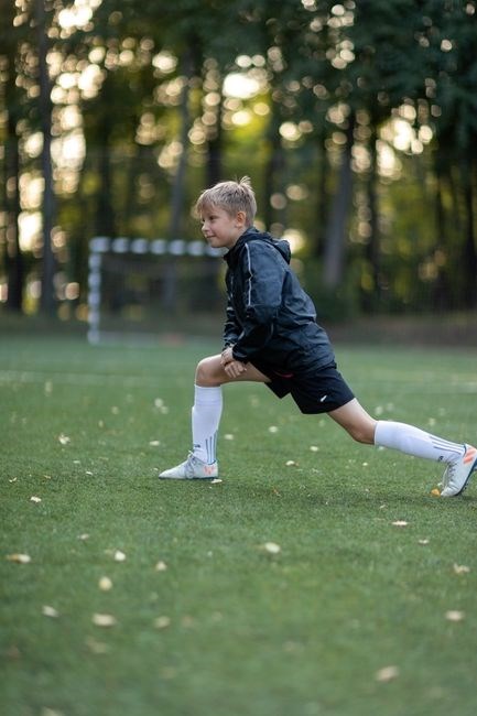
Preventing Severs disease starts with early screening during growth spurts and a consistent warm‑up routine that includes ankle circles, heel‑to‑toe walks, and light calf rolls. Strengthening the calf muscles with step‑up calf raises (2–3 sets of 15 reps) and maintaining flexibility through static stretches held 30–45 seconds, three times daily, builds resilience. Choosing shoes with arch support, a cushioned heel, and a low‑to‑high‑heel‑drop profile reduces impact on the growth plate. When activity increases, a 1–2 cm heel lift can off‑load the apophysis. Education is essential: children should report pain early, avoid over‑training, and rest when discomfort occurs. After pain resolves, a graded return‑to‑sport protocol begins with low‑impact drills for 5–10 minutes, then progresses to sport‑specific movements, each stage lasting 2–3 days before advancing, while continuing calf stretches and ankle mobility drills. If pain recurs, revert to the previous stage and reassess. This combined approach of screening, conditioning, proper footwear, and structured return reduces recurrence risk and supports safe return to play.
Regular monitoring of heel pain during growth spurts, using a simple pain scale, helps clinicians adjust interventions early. Parents should encourage balanced activity, ensuring at least rest day per week, and avoid sudden increases in mileage or intensity. Incorporating foam‑roller calf massages 2–3 times, can circulation and reduce soreness. Finally, a multidisciplinary team—pediatric orthopedists, physical therapists, and coaches—ensures consistent messaging and adherence to the prevention plan!!
PDF Resource Compilation for Clinicians and Patients
Below is a curated list of downloadable PDFs that provide evidence‑based guidance on Severs disease stretches, taping protocols, heel‑lift selection, and orthotic design. Each file is tailored to either a clinician’s practice or a patient’s home program, ensuring clear, actionable steps for prevention and recovery.
- Severs Stretch & Strength Guide – 12‑page PDF detailing calf stretching sequences, toe‑raiser exercises, and closed‑chain dorsiflexion drills with illustrated instructions.
- Taping Techniques Manual – 8‑page handout covering kinesiology tape patterns, application timing, and safety precautions for pediatric patients.
- Heel Lift Selection Sheet – 4‑page worksheet that helps clinicians calculate optimal heel‑lift height based on foot length and pain severity.
- Orthotic Design Blueprint – 10‑page schematic for custom foot orthoses, including growth‑plate support zones and material recommendations.
- Return‑to‑Sport Protocol – 6‑page progression chart that outlines graded activity milestones, monitoring criteria, and red‑flag symptoms.
- Parent & Athlete Education Pack – 5‑page booklet covering daily care, footwear choices, and injury‑prevention tips.
All PDFs are available for free download via the provided links. Clinicians can incorporate them into records or them for handouts, while patients can reference them at home for consistent, evidence‑based self‑management.